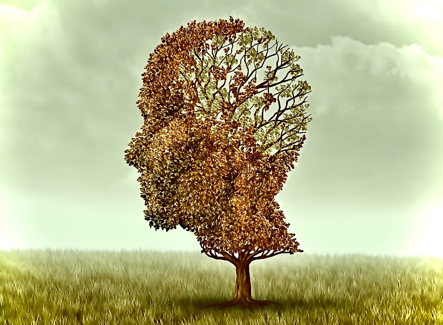
Dementia refers to an impairment of cognitive function, it is a progressive, irreversible clinical syndrome effecting the ability to think, remember, or to make decisions that impacts daily activities 1. It is defined as early-onset (or young-onset) dementia, when the dementia develops before the age of 65 years 2.
The most common cause of late-onset dementia is Alzheimer’s disease (AD), followed by Dementia with Lewy bodies (DLB), vascular dementia (VaD), and frontotemporal dementia (FTD) 3.
The broad term includes a range of symptoms, that may present as difficulties in attention, memory, communication, reasoning, and problem solving, and an atypical decline in vision 4. Many people consider that dementia is an inevitable part of ageing, although despite older age being a most prevalent risk factor, it is not usual that long-term memory and language are affected, in a usual typical ageing process 3. It is unclear as to whether this can be accurately referred to., as an ageing disease.
Causes
There are different types of dementia that are thought to have various causes and known to affect different parts of the brain. This varies the way that it presents as symptoms in the individual. There is also classification for mixed types of dementia, where varied symptoms may present or overlap due to multiple parts of the brain being affected, and with different cause 5.
Types of Dementia
Vascular dementia (VaD) is associated with impaired circulation or blood flow to the brain, resulting in cerebral ischemia or haemorrhagic tissue injury, in specific brain regions 6. VaD has links to metabolic conditions hypertension, diabetes and high cholesterol as potential risk factors 7. Complete mechanisms in the onset of the brain injury are yet to be clearly defined, although it is considered to be a multi-factored accumulation of various events and processes, such as glutamatergic and oxidative stress 8, cerebral ischemia, immune dysfunction 9, calcium overload of neuronal cells 10, augmentation of amyloid peptide, τau proteins and the cholinergic hypothesis 11. Other contributing factors are being considered with increased interest such as inflammation and its role in the occurrence of arteriosclerosis and cerebral vessel disease, that may lead to cerebral infarction 12, 13.
Lewy body dementia (LBD) is defined as neuropathological characteristics of misfolded neuroprotein α-synuclein aggregates in nerve cell bodies and processes, known as Lewy bodies (LB) and Lewy neurites (LN) 14, 15.
LBD shares some of the typical symptoms of dementia such as memory decline, and additionally may also present as some parkinsonian overlap such as extrapyramidal signs, visual hallucinations, changes to alertness, or difficulty in sleeping. There may also be balance and mobility disturbance, or stiffness may occur. It may be relevant that pathological features of LBD, are also often found as a co-pathology in Alzheimer’s Disease 16.
Frontotemporal Dementia occurs as a result of neuronal damage at the frontal and temporal lobes of the brain, that may be due to three subclasses depending on the presence of aggregated proteins., most frequently TAR DNA-binding protein 43 (TDP-43), however τau, and fused in sarcoma (FUS), may also be among these molecular subgroups 17.
The type of dementia itself may be classified into behavioural variant frontotemporal dementia (bvFTD) 18, primary progressive aphasia (PPA), or movement disorders 19. Due to the region of the brain that is affected, there may be some affect to personality and social behaviour, as well as communication in speech, language, writing, and comprehension (aphasia). Neurological disorders may occur as a result of atrophy, (shrinking) of the nerve cells in certain brain regions. This is characteristically progressive, and gradual.
Alzheimer’s disease (AD) is the most frequent type of dementia 20, and similarly to some aspects of FTD and LBD, is widely thought to be characterised by the accumulation of misfolded proteins, known as proteinopathies 21.These proteins can develop as neurofibrillary tangles, made up of looped or twisted τau material, or plaques that form as proteinaceous extracellular deposits consisting of β-amyloid (Aβ) 22.
‘The amyloid hypothesis‘ 23 suggests that an Aβ accumulation in the brain, triggers a cascade resulting in the formation of neurofibrillary tangles via τau protein hyperphosphorylation. Numerous biochemical responses occur as a result, including oxidative stress, excitotoxicity, local inflammation, and cytokine release 24, 25.
The responses influence the structures at the surrounding neuronal cells progressively, and are strongly associated with loss and disruption at the neuron signalling site, known as the synapse. This creates dysfunction at neurotransmitters, and results in neuronal death, and the characteristic cognitive decline seen in individuals with AD 26, 27.
AD is usually associated with short term memory decline that may progressively advance to long-term memory over time. There are thought to be links to genetic factors, providing a 10-30% higher risk factor, if a first degree relative has the condition 28.
Underlying factor & reversible cause: A sub-classification of dementia is identified for where there is an underlying factor, or reversible cause that can be treated 29. These may include chronic infection, thyroid or hormone imbalances, vitamin deficiency, medication side effects, or an increased pressure in the brain, that causes the dementia.
Who can develop dementia?
Although anyone can be affected by dementia, there are increased prevalence in some groups of individuals making them more likely to develop the conditions. The most frequent factor is age, where the majority of individuals known to develop the conditions are aged 65yrs and older 30.
Genetic factors are also thought to be of significant risk. If there is a parental family history (PFH) of the disease, then there is thought to be an average lifetime risk of 15%–25%, as opposed to decreasing to 10%–12% likelihood, where there is no PFH 31.
A Lifestyle for Brain Health (LIBRA) score considered combined risks, when devising tools for individuals’ potential for dementia risk reduction (DRR) 32.
These include Individuals with existing heart related conditions including unmanaged hypertension, high cholesterol and smoking, that are all known to increase likelihood of vascular and other forms of dementia, whilst traumatic brain injury [particularly where it is severe, prolonged or reoccurring], may also significantly increase risks of developing onset of the dementia diseases 32.
Treatment
Research is ongoing toward preventing the onset of cognitive decline, and for new lines as potential treatments of the diseases, however there is currently no cure for dementia 3. Treatment aims remain to slow down the progression of the disease.
Potential drug targets and Pharmacotherapy
Treatment options available will depend on the type of dementia diagnosed, presentation of the condition, and stage of disease progression.
There is no approved drug to address either Frontotemporal Lobe or Lewy Body Dementia. In FTD, it is likely that main objectives of therapeutic targets, will be in preventing protein misfolding and aggregation, or in aggregate clearing. Many studies continue to find little evidence in support of lines that focus on pathways to recover loss of function 33. There have been various drugs that have shown some disease-modifying potential for LBD 34. Formerly, studies have focused on vaccination as a potential treatment for synucleinopathies 35, whilst antibodies against α-synuclein have also been developed, finding some effects of immunotherapies against α-synuclein. However clinical data remains inconclusive, and is sparse in human studies 36, 37.
Over the last 10 years there has been little advancement in pharmacological therapies for AD 38.
One of the most frequent targets for drug therapies in the treatment of AD symptoms 3, aims to preserve the neurotransmitter acetylcholine (ACh) levels at the synaptic cleft. This is because, decreased biosynthesis of ACh, characterises the earlier stages of AD progression. Therefore, drug strategies such as acetylcholinesterase inhibitors (AChEIs) (e.g. donepezil, galantamine and rivastigmine), act by inhibiting the cholinesterase enzyme that would usually be breaking down the ACh molecules 39.
The N-methyl-D-aspartate (NMDA) receptor, is a receptor of the primary excitatory neurotransmitter in the brain, glutamate. It has a significant position in AD development., when stimulated results in an influx of calcium ions (Ca2+), that triggers a signalling pathway, activating genes that are involved in the formation of long-term potentiation (LTP), or the strengthening of synapses 40. When the stimulation of the NMDAR’s and its higher influx of Ca2+ is excessive, the result is dysfunction at the synapse, excitotoxic damage, apoptotic cell death, and thus cognitive decline 41. Therefore, an antagonistic targeting of this receptors’ activation., holds a significant position in AD management (e.g. memantine for severe Alzheimer’s disease and vascular dementia) 3.
Characterised fibrillary formations in AD, are thought to occur due to an imbalanced protein homeostasis mechanism within the cells, and it is soluble-amyloid oligomers and τau molecules that are thought to be the significant neurotoxic mediators of the disease 42. Where this solubility has been associated with the increased toxic effects, a potential target for treatment options may be in converting the soluble protein into an insoluble form at adduct formation, thus reducing the toxicity response 43.
A susceptibility to Aβ aggregation, is known to be significantly higher with an oxidative stress response on association with Zn2+, Fe2+ and Cu 2+ metal ions 44. Some protective response from polyphenol groups (as we shall see inPart II) have been associated with metal chelation processes 45.
Disordered proteins that usually arise with formation of fibrillary tangles, usually trigger a response known as the unfolded protein response (UPR), and this has been shown to be dysfunctional in Alzheimer’s conditions 46. Respectively, initiating the UPR response, is shown to improve the symptoms that are characteristic of AD 47. One strategy in directing potential treatments, may be to maintain homeostasis by facilitating the activation of the UPR.
Symptom Management.
Drug treatments that are non-specific to dementia, are usually prescribed for behavioural and psychological symptoms of dementia (BPSD). 90% of people with dementia experience some form of neuropsychiatric symptoms including anxiety, depression, apathy, insomnia, agitation or hallucination 27.
Belsomra (Suvorexant), an Orexin Receptor Antagonist (ORA) medication usually approved outside of Europe for symptoms of dementia-related insomnia, was recently studied for approval in the UK (October 2023) 48. The treatment works by antagonising the neurotransmitter orexin receptor 49. Orexin has a fundamental role in the sleep-wake cycle regulation, maintaining wakefulness, and has been closely associated with a role in both cognitive function, and in the pathology of AD 50. Secretion of orexin mostly occurs at the lateral hypothalamus, then binding to the G-protein–coupled receptors (GPCRs), and activating an alternative downstream signalling pathway, other than the classic pathway 51, 52.
Drug Development and Relevance of Repositioning.
Amongst clinical research for therapeutic interventions toward dementia, a strategy of repositioning existing drug therapies has been a frequent consideration 53. This involves identifying of a medication that is shown to be effective to treat one condition, then using it to treat a different condition 54.
An example of this, are the drug groups usually indicated for use in hypertension., due in part to an associated incidence of midlife hypertension being a frequent earlier indication of vascular dysregulation associated with dementia development later in life 55, and also due to some existing evidence that by blocking the angiotensin II receptor and calcium channel signalling in particularly., some antihypertensive groups have shown some evidence of neurological benefit 56.
In this way, it may then be hypothesised that this drug group may also have a positive effect on neuronal plasticity for various neurodegenerative diseases, and possess neuroprotective qualities for studies of dementia. A cumulative 124 studies between 2012 and 2022, suggested a potential 573 drugs for AD repurposing candidates 57.
Drug candidates for repositioning in the case of dementia or AD, may then give rise to translation of priority compounds for taking forward to clinical trials, based on consensus, or formally established drugs and their proposed mechanisms of action 53. As an example, 6 pharmacological categories of drug mechanisms that are considered when identifying candidates for the repositioning into use for dementia and Alzheimer’s Disease., may be suggested as follows, anti-hypertensive, anti-diabetic, neurologic, antibiotic, anti-inflammatory or anticancer 58.
Finally, this subject may introduce a topic of qualities that may be considered when devising non-pharmacological approaches to the treatment of dementia, and in particularly where these properties may be made accessible and available through diet and nutrition, when devising functional food strategies for individuals who have been diagnosed with dementia.
5. Custodio, N., Montesinos, R., Lira, D., Herrera-Pérez, E., Bardales, Y. & Valeriano-Lorenzo, L. (2017) Mixed dementia: A review of the evidence. Dement Neuropsychol. 2017 Oct-Dec;11(4):364-370.
6. Sanders, A. E., Schoo, C. & Kalish, V. B. (2023) Vascular Dementia. StatPearls [Internet]. StatPearls Publishing; Treasure Island (FL): Oct 22, 2023.
7. O’Brien, J. T. & Thomas, A. (2015) Vascular dementia. Lancet. 2015 Oct 24;386(10004):1698-706.
8. Busse, M., Kunschmann, R., Dobrowolny, H., Hoffmann, J., Bogerts, B., Steiner, J., et al. (2018) Dysfunction of the blood-cerebrospinal fluid-barrier and N-methyl-D-aspartate glutamate receptor antibodies in dementias. Eur Arch Psychiatry Clin Neurosci. 2018;268(5):483–492.
9. Mosek, A., Yust, I., Treves, T.A., Vardinon, N., Korczyn, A. D. & Chapman, J. (2000) Dementia and antiphospholipid antibodies. Dement Geriatr Cogn Disord, 11 (2000), pp. 36-38
10. Raza, M., Deshpande, L. S., Blair, R. E., Carter, D. S., Sombati, S. & DeLorenzo, R. J. (2007) Aging is associated with elevated intracellular calcium levels and altered calcium homeostatic mechanisms in hippocampal neurons. Neuroscience Letters. 2007;418(1):77–81.
11. Román, G. C. & Kalaria, R. N. (2006) Vascular determinants of cholinergic deficits in Alzheimer’s disease and vascular dementia. Neurobiol Aging. 2006 Dec;27(12):1769-85.
12. Bir, S. C., Khan, M.W., Javalkar, V., Toledo, E.G., Kelley, R.E. (2021) Emerging Concepts in Vascular Dementia: A Review. J. Stroke Cerebrovasc. Dis. 2021; 30:105864.
13. Kalaria, R.N. (2016) Neuropathological Diagnosis of Vascular Cognitive Impairment and Vascular Dementia with Implications for Alzheimer’s Disease. Acta Neuropathol. 2016; 131:659–685.
14. Taylor, J. P. et al. (2020) New evidence on the management of Lewy body dementia. Lancet Neurol. 19, 157–169 (2020).
15. Hansson, O. (2021) Biomarkers for neurodegenerative diseases. Nat. Med. 27, 954–963 (2021).
16. Herrmann, F. R., Burkhard, P. R., Bouras, C. & Kovari, E. (2013) Neuropathology of dementia in a large cohort of patients with Parkinson’s disease. Parkinsonism Relat. Disord.19, 864–868.
17. Scarioni, M., Gami-Patel, P., Timar, Y., Seelaar, H., van Swieten, J. C., Rozemuller, A. J. M., et al. (2020) Frontotemporal Dementia: Correlations between Psychiatric symptoms and Pathology. Ann Neurol 87(6):950–961
18. Kumar Chanchal, D., Singh, K., Bhushan, B., Singh Chaudhary, S., Kumar, S., Kumar Varma, A., Agnihotri, N. & Garg, A. (2023) An updated review of Chinese skullcap (Scutellaria baicalensis): Emphasis on phytochemical constituents and pharmacological attributes. Pharmacological Research – Modern Chinese Medicine, Vol. 9,2023,100326, ISSN 2667-1425.
19. Forman, M. S., et al (2006) Frontotemporal dementia: clinicopathological correlations. Ann Neurol 59(6):952–962
20. Reitz, C. & Mayeux, R. (2014) Alzheimer disease: epidemiology, diagnostic criteria, risk factors and biomarkers. Biochem Pharmacol. 2014 Apr 15;88(4):640-51.
21. Allegri, R. F. (2020) Moving from neurodegenerative dementias, to cognitive proteinopathies, replacing “where” by “what”…. Dement Neuropsychol. 2020 Jul-Sep;14(3):237-242.
22. Hardy, J. & Selkoe, D. J. (2002) The amyloid hypothesis of Alzheimer’s disease: Progress and problems on the road to therapeutics. Science 2002, 297, 353–356.
23. Hardy, J. (2017) The discovery of Alzheimer-causing mutations in the APP gene and the formulation of the “amyloid cascade hypothesis”. FEBS J. 2017, 284, 1040–1044.
24. Yin, X., Qiu, Y., Zhao, C., Zhou, Z., Bao, J., & Qian, W. (2021) The Role of Amyloid-Beta and Tau in the Early Pathogenesis of Alzheimer’s Disease. Med. Sci. Monit. 2021;27: e933084.
25. Baazaoui, N., & Iqbal, K. (2018) A Novel Therapeutic Approach to Treat Alzheimer’s Disease by Neurotrophic Support During the Period of Synaptic Compensation. J. Alzheimer’s Dis. 2018; 62:1211–1218.
26. Montero-Crespo, M., Domínguez-Álvaro, M., Alonso-Nanclares, L., DeFelipe, J., & Blazquez-Llorca, L. (2021) Three-dimensional analysis of synaptic organization in the hippocampal CA1 field in Alzheimer’s disease. Brain. 2021; 144:553–573.
27. Ferrer, I. (2009) Altered mitochondria, energy metabolism, voltage-dependent anion channel, and lipid rafts converge to exhaust neurons in Alzheimer’s disease. J. Bioenerg. Biomembr. 2009; 41:425–431.
28. Schachter, A. S. & Davis, K. L. (2000) Alzheimer’s disease. Dialogues Clin Neurosci. 2000 Jun ;2(2) :91-100.
29. Tripathi, M. & Vibha, D. (2009) Reversible dementias. Indian J Psychiatry. 2009 Jan;51 Suppl 1(Suppl1): S52-5. PMID: 21416018; PMCID: PMC3038529.
31. Goldman, J. S., Hahn, S. E., Catania, J. W., et al. (2011) Genetic counselling and testing for Alzheimer disease: joint practice guidelines of the American College of medical genetics and the National Society of genetic Counselors. Genet Med 2011;13:597–605.
32. Deckers, K., van Boxtel, M. P. J., Schiepers, O. J. G., et al. (2015) Target risk factors for dementia prevention: a systematic review and Delphi consensus study on the evidence from observational studies. Int J Geriatr Psychiatry 2015; 30:234–46. 10.1002/gps.4245
33. Cerejeira, J., Lagarto, L. & Mukaetova-Ladinska, E. B. (2012) Behavioral and Psychological Symptoms of Dementia. Front. Neurol. 2012; 3:73.
34. Turner, R.S., Hebron, M.L., Lawler, A., Mundel, E. E., Yusuf, N., et al. (2020) Nilotinib Effects on Safety, Tolerability, and Biomarkers in Alzheimer’s Disease. Ann Neurol. 2020 Jul;88(1):183-194.
35. Brown, D.G., Shorter, J. & Wobst, H.J. (2020) Emerging Small-Molecule Therapeutic Approaches for Amyotrophic Lateral Sclerosis and Frontotemporal Dementia. Bioorg. Med. Chem. Lett. 2020; 30:126942.
36. George, S. & Brundin, P. (2015) Immunotherapy in Parkinson’s Disease: Micromanaging Alpha-Synuclein Aggregation. J. Park. Dis. 2015; 5:413–424.
37. Boxer, A.L., Lang, A.E, Grossman, M., Knopman, D.S., Miller, B.L., Schneider, L. S. et al. (2014). Davunetide in patients with progressive supranuclear palsy: a randomised, double-blind, placebo-controlled phase 2/3 trial. Lancet Neurol. 2014 Jul;13(7):676-85. Epub 2014 May 27.
38. Cummings, J. L., Tong, G., & Ballard, C. (2019) Treatment Combinations for Alzheimer’s Disease: Current and Future Pharmacotherapy Options. J. Alzheimer’s Dis. 2019; 67:779–794.
39. Breijyeh, Z. & Kara-man, R. (2020) Comprehensive Review on Alzheimer’s Disease: Causes and Treatment. Molecules. 2020; 25:5789.
40. Wang, R., & Reddy, P.H. (2017) Role of Glutamate and NMDA Receptors in Alzheimer’s Disease. J. Alzheimer’s Dis. 2017; 57:1041111–1048.
41. Briggs, R., Kennelly, S.P. & O’Neill, D. (2016) Drug treatments in Alzheimer’s disease. Clin. Med. 2016; 16:24777777–253.
42. Bi, X. (2010) Alzheimer disease: Update on basic mechanisms. J. Am. Osteopat. Assoc. 2010;110: S3–S9.
43. Ladiwala, A. R. A., Lin, J. C., Bale, S. S., Marcelino-Cruz, A. M., et al. (2010) Resveratrol Selectively Remodels Soluble Oligomers and Fibrils of Amyloid Aβ into Off-pathway Conformers. J. Biol. Chem. 2010; 285:24228–24237.
44. Curtain, C. C., Ali, F., Volitakis, I., Cherny, R.A., Norton, R. S., Beyreuther, K., Barrow, C.J., Masters, C.L., Bush, A.I., & Barnham, K.J. (2001) Alzheimer’s Disease Amyloid-β Binds Copper and Zinc to Generate an Allosterically Ordered Membrane-penetrating Structure Containing Superoxide Dismutase-like Subunits. J. Biol. Chem. 2001; 276:204666–20473.
45. Mandel, S. A., Amit, T., Weinreb, O. & Youdim, M.B. (2011) Understanding the Broad-Spectrum Neuroprotective Action Profile of Green Tea Polyphenols in Aging and Neurodegenerative Diseases. J. Alzheimer’s Dis. 2011; 25:1877–208.
46. Hetz, C., Chevet, E. & Harding H.P. (2013) Targeting the unfolded protein response in disease. Nat. Rev. Drug Discov. 2013; 12:703–719.
47. Cornejo, V. H., Hetz, C. (2013) The unfolded protein response in Alzheimer’s disease. Semin. Immunopathol. 2013; 35:27–292.
49. Muehlan, C., Roch, C., Vaillant, C., & Dingemanse, J. (2023). The orexin story and orexin receptor antagonists for the treatment of insomnia. Journal of Sleep Research, 32(6), e13902.
50. Um, Y.H. & Lim H.K. (2020) Orexin and Alzheimer’s Disease: A New Perspective. Psychiatry Investig. 2020; 17:616.
51. Wang, C., Wang, Q., Ji, B., Pan, Y., Xu, C., Cheng, B., Bai, B., Chen, J. (2018) The Orexin/Receptor System: Molecular Mechanism and Therapeutic Potential for Neurological Diseases. Front. Mol. Neurosci. 2018; 11:220.
52. Health Research Authority (2023) A Phase III Randomized, Placebo-Controlled Clinical Trial to Study the Safety and Efficacy of Suvorexant (MK-4305) for the Treatment of Insomnia in Subjects with Alzheimer’s Disease. Online: ttps://www.hra.nhs.uk/planning-and-improving-research/application-summaries/research-summaries/safetyefficacy-of-suvorexant-for-treatment-of-insomnia-in-ad-subjects/. Accessed: 20 April 2024.
53. Corbett, A. et al. (2012) Drug repositioning for Alzheimer’s disease. Nat. Rev. Drug Discov 11, 833–846 (2012).
54. Sirota, M., Dudley, J. T., Kim, J., Chiang, A. P., Morgan, A. A., Sweet-Cordero, A., Sage, J. & Butte, A. J. (2011) Discovery and preclinical validation of drug indications using compendia of public gene expression data. Sci Transl Med, 3(96), 96ra77
55. Kehoe, P. G., et al. (2009) Angiotensins in Alzheimer’s disease – friend or foe? Trends Neurosci. 2009 Dec;32(12):619–28.
56. Kehoe, P. G. & Passmore, P. A. (2012) The renin-angiotensin system and antihypertensive drugs in Alzheimer’s disease: current standing of the angiotensin hypothesis? J Alzheimers Dis JAD. 2012;30 Suppl 2: S251–268.
57. Grabowska, M. E., Huang, A., Wen, Z., Li, B. & Wei, W. Q. (2023) Drug repurposing for Alzheimer’s disease from 2012-2022-a 10-year literature review. Front Pharmacol. 2023 Sep 7; 14:1257700.
58. Zagórska, A., Czopek, A., Fryc, M., Jaromin, A. & Boyd, B. J. (2023) Drug Discovery and Development Targeting Dementia. Pharmaceuticals (Basel). 2023 Jan 19;16(2):151.
Botanical Information: World Flora Online (WFO) http://www.worldfloraonline.org. Accessed: June 2024. and POWO (2026). “Plants of the World Online. Facilitated by the Royal Botanic Gardens, Kew. Published on the Internet; https://powo.science.kew.org/.
Originally published: 24/06/2024 This article was recently reviewed by author for accuracy and updated on 2nd October, 2026 to include the latest information and taxonomy.
Whilst every care is taken to ensure accurate, reputable and the most recent sources and research for this information, this content is not intended as substitute for advice or treatment from a professional clinician. As with conventional medicines, herbal and natural medicines have active ingredients and can have potent effects and interactions with the body, medications, and with other conditions. Especially for those who are taking prescription or over-the-counter medications, it is advised to consult a professional herbalist or medical doctor before beginning any herbal, supplement or lifestyle program. Care and responsible administration is always strongly advocated.
Apothēcary of Botanical Medicine
South Westminster Centre For Health, 82 Vincent Square, London SW1P 2PF